Optopol

No longer available
Optopol
€55,900 (EUR)
Location:Aalten, Netherlands
Description
This is a Demo.
Dat mean he is not used but he is also not in the packing as new.
Thats why also the price is cheaper. We may not also to sale it to all countries. We have first to get a permission.
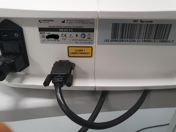
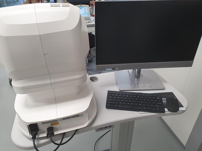
OPTICAL COHERENCE TOMOGRAPHY
REVO FC with 80 000 A-scan/sec scanning speed offers advanced technologies and remarkable simplicity of operation. It meets all requirements for modern optical tomographs.
RETINA
Single 3D Retina examination is enough to perform both Retina and Glaucoma analysis based on retinal scans. Software automatically recognizes 8 retina layers. Thus allowing a more precise diagnosis and mapping of any changes in the patient’s retina condition.
GLAUCOMA
Comprehensive glaucoma analytical tools for quantification of the Nerve Fiber Layer, Ganglion layer and Optic Head with DDLS allow for the precise diagnosis and monitoring of glaucoma over time.
With the golden standard 14 optic nerve parameters and a new Rim to Disc and Rim Absence the description of ONH condition is quick and precise.
Advanced view which provides combined information from Retina and Disc scan to integrate details of the Ganglion cells, RNFL, ONH in a wide field perspective for comprehensive analysis.
Advance Retina & ONH
ONH Single
GLAUCOMA Advance Retina & ONH GLAUCOMA ONH Single
Asymmetry Analysis of Ganglion layers between hemi-spheres and between eyes allows easier identification and detection of glaucoma in early stages and in non-typical patients.
Ganglion Both
Ganglion Progression
GLAUCOMA Ganglion Both GLAUCOMA Ganglion Progression
Implemented the DDLS - Disc Damage Likelihood Scale which use 3 separate classification for small, average and large discs. It supports the practitioners in a quick and precise evaluation of the patient’s glaucomatous disc damages.
ONH Both
ONH Progression
GLAUCOMA ONH Both GLAUCOMA ONH Progression
COMPLET YOUR GLAUCOMA REPORT
To eliminate common problem with the understanding of the patient’s IOP pachymetry module provides IOP Correction value. With the implemented Adjusted IOP formula you can quickly and precisely understand the measured IOP value.
As the Pachymetry and Anterior Chamber Angle Verification require no additional attachments, the predefined Glaucoma protocol, which consists of Retina, Disc and Anterior scans, can be done automatically to reduce patient chair time.
Closing angle
Anterior single view
Closing angle Anterior single view
* Images courtesy of Prof. Edward Wylęgała MD, PhD
COMPREHENSIVE GLAUCOMA SOLUTION
STRUCTURE & FUNCTION - Combined OCT and VF results analysis
Invaluable combination of information about the functional quality of vision with comprehensive data on retinal Ganglion Cells, RNFL and Optic Nerve Head for both eyes on a single report page. The S&F report contains the following:
VF sensitivity results (24-2/30-2 or 10-2)
Total and Pattern Deviation probability graphs for VF results
Reliability and Global indices for VF results
Combined map of Structure & Function
Ganglion cell analysis (GCL+IPL or NFL+GCL+IPL)
ONH and NFL analysis including charts and comparison tables
NFL Asymmetry Plot
The S&F report compares in a natural way the anatomical relationship between VF and RNFL/Ganglion maps.
Structure & Function
STRUCTURE & FUNCTION - Combined OCT and VF results analysis
SINGLE PAGE REPORT
S+F provides a quick and comprehensive single page report for glaucoma management.
ANTERIOR
Anterior segment lens for standard and wide examinations is built into the device. No additional lens is required.
Cornea single
Cornea both
ANTERIOR Cornea single ANTERIOR Cornea both
Cornea Comparison Cornea Progression
Anterior Cornea Comparison Anterior Cornea Progression
FOLLOW UP
High density of standard 3D scan allows to precisely track the disease progression. Operator can analyze changes is morphology, quantified progression maps and evaluate the progression trends.
Progression Morphology Progression Quantification
FOLLOW UP Progression Morphology FOLLOW UP Progression Quantification
COMPARISON
Comparison view is an easy and convenient way to review results from two separate visits. The view is available for most of the scan programs.
NETWORKING
A proficient networking solution increases productivity and an enhanced patient experience. It allows you to view and manage multiple examinations from review stations in your practice. Effortlessly helping to facilitate patient education by allowing you to interactively show examination results to patients. Every practice will have different requirements which we can provide by tailoring a bespoke service. There is no additional charge for the server module.
DICOM
Store, exchange, and transmit results through DICOM gateway to the hospital network.LEARN MORE ABOUT OPTIONAL SOFTWARE MODULES
ANGIOGRAPHY OCT This module allows visualization of the retinal microvasculature. Angiography SOCT is a non-invasive, dye-free technique providing 3D image of retinal blood circulation.
BIOMETRY OCT
B-OCT™ innovative method of using the posterior OCT device to measure ocular structure along eye axis.TOPOGRAPHY OCT1
T-OCT™ is a pioneering way to provide detailed corneal Curvature maps by using posterior dedicated OCT.
Specifications
| Manufacturer | optopol |
| Model | Revo FC |
| Year | 2019 |
| Condition | New |